PREOPERATIVE PLANNING – ORTHOPEDIC SURGERY
In orthopedic surgery, preoperative planning is an essential step and an effective way for surgeons to anticipate potential surgical challenges and begin targeting the choice of implants. However, in order to ensure the best possible solution, they must take into account numerous criteria, including the patient’s clinical presentation, type of procedure, choice of prosthesis model, and prosthesis placement…
In many cases, the specific anatomical characteristics of patients require them to order multiple prostheses to ensure they have the appropriate materials during the operation. However, this operating method poses several problems:
How can we limit costs and optimize the management of medical resources? How can we ensure that the surgical technique and implant are as effective as possible?

We have developed a 3D reconstruction and anatomical landmark detection software for ankles, hips, and knees. This solution allows surgeons to precisely visualize the patient’s anatomy preoperatively in order to establish the necessary surgical procedures.
Surgeons benefit from a technology that saves them time and provides precision in the planning of their surgical procedures.
Neovision has developed a solution that enables 3D reconstruction of the bones of the lower limb and anatomical landmark detection. We have leveraged a deep learning algorithm and standard DICOM medical imaging data (a series of stacked 2D cross-sectional images that create a volume).
3D Reconstruction
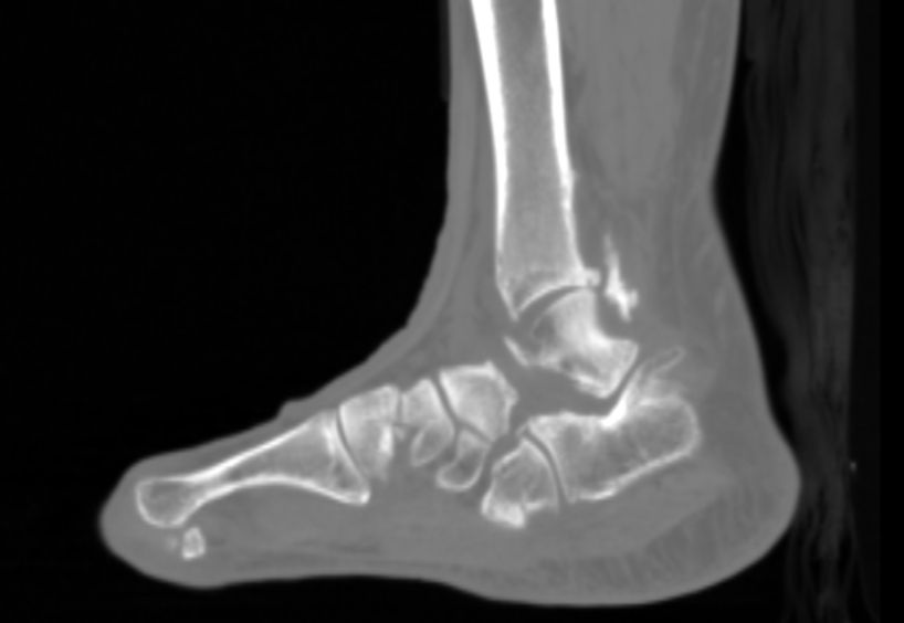
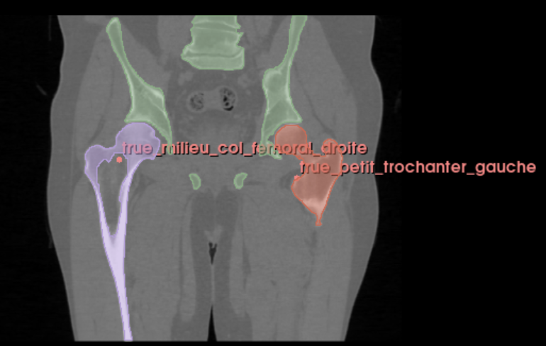
This functionality automates the task of manual reconstruction performed by medical specialists by utilizing an algorithm instead. This automated process addresses a 3D segmentation challenge, precisely obtaining the bones of the lower limb (pelvis, femur, tibia), especially the joint areas that are complex and crucial for surgeons. To accomplish this, we transformed the DICOM data into Nifti format, which is suitable for deep learning tasks in 3D. The segmentation representation is a point cloud that describes the bone surface and defines the contours of each bone.
Landmarking
Neovision has developed a landmarking algorithm, which involves the identification of strategic anatomical points for each bone (such as the tibial plateau, talar dome in the case of the ankle, for example). To achieve this, we relied on the knowledge of orthopedic surgeons to create a database of anatomical points that are essential for successful surgery.
The combination of these two approaches enables surgeons to assess, prior to the operation, what the most optimal surgical decision will be. This includes selecting the appropriate prosthesis, determining its positioning, and planning the bone cuts
18 July 2023
Computer Vision, Deep Learning, Imagerie 3D, R&D, Santé